Diary Of A Cancer Researcher
  • Home
  • About
  • Blog
  • External Work
  • Gallery
  • Social Media
  • Further Info
  • FAQ
  • Home
  • About
  • Blog
  • External Work
  • Gallery
  • Social Media
  • Further Info
  • FAQ

Blog...

What the Hell? - Cancer Part 2

5/7/2017

0 Comments

 
In this edition of "What the Hell? - Cancer" I will going through how cancers are diagnosed. This will hopefully give you an idea about what the different stages of cancer are and how each cancer is graded differently. 

NOTE: All my examples will be using breast cancer because it is the cancer type I study and am most familiar with.

Before I begin I want to just clear up the differences between primary tumours, secondary tumours and primary secondary tumours. 
  • Primary tumour: tumour which has originated in one location 
  • Secondary tumour: tumour which is derived from the primary tumour (share genetic etc. similarities) but is found in one or more locations in different locations in the body from the primary tumour (happens usually by metastasis).
  • Primary secondary tumour: tumour which has originated independently from another tumour in a different part of the body at a different time to the other tumour. E.g a breast cancer patient is discovered to have colorectal cancer (CRC) after diagnosis with breast cancer however the tumours are not related (genetically etc.). The CRC tumour developed independently from the breast tumour. 
Cancer diagnosis is a tricky business. If you read my awareness posts you will see the signs and symptoms of a lot of different cancers. You may have noticed that a lot of the signs and symptoms are generic for example bloating, intestinal discomfort, a prolonged cough etc.

Detection

Tumours need to be detected before you can take a biopsy etc. These are the main ways tumours are detected (but not the only ways). Apologies for the explanations. While I learnt all this in my masters (woo nuclear physics) I am no physicist... 
  • Ultrasound: this technique is based on sound waves. Sound wave are sent into the body and the speed the sound waves move tells you what type of tissue is there. The slower the sound waves the denser the tissue (e.g bone 4080 m/s vs soft tissue 1540 m/s and air 330 m/s). The sound also bounces back from the tissue. The denser the tissue (e.g bone) the more sound is sent back.  An ultrasound machine sends out a pulse which is reflected back to the machine. Depending on the speed of the reflection and how much sound is reflected back this creates an "image". Tumours are denser than normal soft tissue.
 
  • PET/CT: this techniques uses a modified type of glucose (18FDG) that comes up black on a PET scan. As tumours use a huge amount of glucose you can infer that places where there are dark areas of glucose are masses of highly proliferating cells. A PET scan takes an image of the body however the resolution is quite low, meaning you are not 100% sure where exactly in the body the tumour is. A CT scan is a much more in depth image of the body, including the organs etc. by fusing the images you get a picture of where in the body this build up of glucose is and where the tumour is. There is one drawback with this method - the bladder. The bladder is where the modified glucose goes to be excreted and therefore you will always see the bladder as a big black spot. This can hinder diagnosing bladder cancer and cancers around that area. 
 
  • MRI (magnetic resonance imaging): this technique is based on creating a magnetic field. Interestingly, MRIs look mainly at water molecules. Two magnetic fields are created perpendicular to each other. The first magnetic field forces hydrogen nuclei to align parallel (low energy state) or anti-parallel (high energy state) to the magnetic field. Each tissue type has a certain number of high energy and low energy nuclei. The difference between the two gives each tissue type an "M" value. For example if a tissue had 20 low energy nuclei and 100 high energy nuclei the M Value would be 5 (100/20). The low energy nuclei are then "excited" to high energy nuclei by adding energy. As they return to a low energy state, they release energy. The MRI detects how much energy is released. Different tissues release different amounts of energy, allowing the machine to tell them apart. The second magnetic field allows the MRI to create an image. The MRI does all of this in small sections and creates a picture by building the sections one on top of the other. Nuclear physics everyone! 

Pathology

When you perform a biopsy you take a small piece of the tumour to figure out what type of tumour it is and how severe it is. This gives you the best idea to treat the tumour. 

The first step after taking a biopsy is to store it. Usually tissue is fixed in formalin or flash frozen. The tissue is then sent to a pathologist who embeds it in a wax block. This allows the pathologist to cut tiny slices from the tumour (I mean micron sizes, barely the width of a single hair). The wax is removed and the tissue is stained. The staining allows you to see normal cells, cancer cells and blood vessels. You can also use this method with antibodies which bind to proteins on the outer surface of the cells for example Ki67 which tells you how fast the cells are growing. 
Picture
Staining of mice lung cancer tissue. The large purple clumps of cells are cancerous tissue.
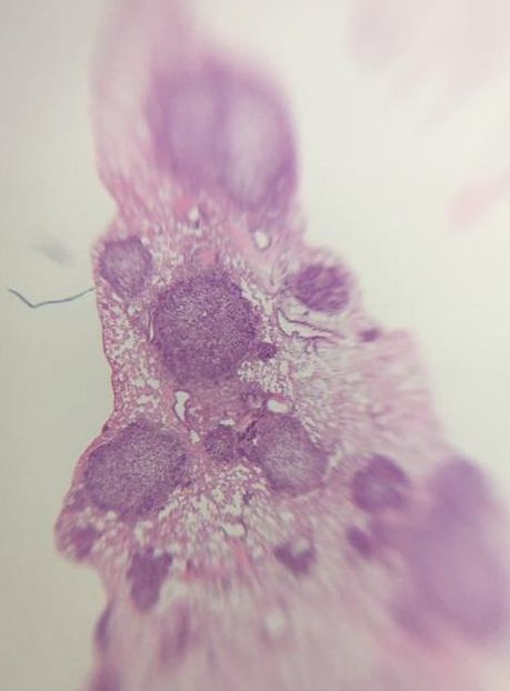
Part of what pathologists are looking for when they stain biopsies is structural changes to cells. Cells "adapt" to their environment or from internal cues by changing their structure. There are a number of cellular adaptions which happen normally but can also be due to disease etc.
  1. Atrophy: this is when the cells shrink. This can happen in response to not using them (e.g. muscle) or poor blood supply/nutritional deficiency. 
  2. Hypertrophy: this is where the number of cells in an organ or tissue increase, mostly seen in skeletal muscle and cardiac muscle. 

The next three are characteristic of pre-cancer cells (i.e. structural changes that can lead to cancer):
  1. Hyperplasia: number of cells in organ or tissue increases (similar to hypertrophy).
  2. Metaplasia: cells change from one type to another. For example in your oesophagus you have epithelial cells (roughly circular blobs) whereas in you stomach you have mucosal cells which are more rectangular and structured to withstand stomach acids. In Barrett's oesophagus, constant exposure of the oesophagus cells to stomach acid due to acid reflux changes the cells from epithelial to mucosal. Barrett's oesophagus is a risk factor for oesophageal cancer. 
  3. Dysplasia: normal cells grow in a disordered way. Instead of growing side by side where all the cells are orientated the same direction, cells grow all over the place.

Change of shape and structure and the number of darkly stained cells allow pathologists to grade tissue samples. Histopathology is based very much on the person and takes years and years to generate enough knowledge to be able to look down a microscope at a purple stained piece of tissue and say "that is a squamous cell carcinoma" (type of skin cancer - don't look up images). There is a lot more to the work of a pathologist but I don't have the knowledge to delve deeper. ​

Staging and Grading

So what do I mean by stage? Well the stage of the cancer tells you (in general terms) how big the tumour is, if it has invaded locally or systemically and what treatment you should use. 
  • Benign: a collection of cells that do not display the hallmarks of cancer and are not likely to grow and invade
  • Stage 1: this is the earliest stage of a tumour. The tumour is contained within one area and has not invaded. The tumour also tends to be small in comparison to later stages. 
  • Stage 2: the tumour begins to grow and invade into the immediate area around the tumour. 
  • Stage 3: the tumour invades locally and is large
  • Stage 4: the tumour invades systemically (metastasis) 

Clinicians expand on this and use a system called TNM. This is "Tumour", "Node", "Metastasis". 
  • Tumour: numbered 1-4 depending on the tumour stage, the size of the tumour and how much it has spread
  • Node: numbered 1-3 depending on is lymph nodes are involved, size, location and how many are involved.
  • Metastasis: 0 or 1, either there is metastasis or no metastasis
Picture
Figure 1: Generic stages of cancer progression
As I said before each cancer type has slightly different staging criteria. This is due to the type of organ it is in (some organs are smaller than others so a "small" tumour in a large organ is actually quite a big tumour in a smaller organ). The staging also has to take into account where the tumour invades locally and systemically and if lymph nodes are involved. 
​
In breast cancer:
  • Stage 1 (T1): is a tumour which is less than 2cm across and a few breast cells are found in the lymph nodes around the breast.
  • Stage 2 (T2): is a tumour between 2cm and 5cm across and breast cells are found in 1-3 lymph nodes in the armpit/near breastbone
  • Stage 3 (T3): is a tumour greater than 5cm across, has invaded locally (into the chest cavity or skin) and breast cells are found in 4-9 lymph nodes in the armpit/near breastbone
  • Stage 4 (T4) or advanced cancer: is a tumour which has spread systemically (into the brain, liver, bones etc.)

Other cancers like Colorectal Cancer use additional staging techniques for example Duke's Staging which is very like the numbered system but is marked by grade A-D. 

Location/Origin

But the above is not the only way to identify the type of cancer! Oh no, like all aspects of cancer biology there is always more. 

While the stage/grade will help you with treatment options there are other factors such as location that tell you about the cancer.

For example in breast cancer:

Epithelial Tumours:
  • Ductal carcinoma in situ (DCIS) - tumours in the ducts of the breast, non-invasive
  • Lobular carcinoma in situ (LCIS) - tumours in the lobes of the breast, non-invasive
  • Invasive ductal breast cancer - invasive cancer (invades into the basement membrane) originating in the ducts of the breast
  • Invasive lobular breast cancer - invasive cancer (invades into the basement membrane) originating in the lobes of the breast

Other:
  • Angiosarcoma of the breast - tumour arising from the lining of the blood vessels in the breast
  • Paget's disease - cancer of the nipple

Mutations and Chromosomal Changes

And as always we come back to DNA. There are mutations that are common in pretty much every cancer type (e.g. p53) but other mutations are specific to certain cancers of families of cancers. For example BRCA mutations are associated with both breast and ovarian cancers. 

In breast cancer the common mutations used to define cancer types are:
  • Oestrogen receptor positive (ER+)
    • Luminal A - ER+, PR+ 
    • Luminal B - ER+, PR+/-
  • Oestrogen receptor negative (ER-)
  • Her2+
  • Triple negative breast cancer (a collection of tumours with no mutations in ER, PR or Her2+)

In breast cancer if you have suspected Her2+ cancer a number of tests are done such as Immunohistochemistry (IHC) which detects Her2 receptor levels on the surface of the breast cancer cell and Fluorescence In Situ Hybridisation (FISH) which detects the number of copies of the HER2 gene there are.  

There are genetic screening tests used in the clinic for some cancers. In breast cancer there is OncotypeDx which detects 21 genes commonly mutated in breast cancer. This gives you a score. OncotypeDx also has systems for prostate and colon cancer.

Changes to the chromosomes themselves are also common, usually in haematological malignancies.
  • Translocations: where one part of a chromosome switches with another part of the same or different chromosomes
  • Deletions: where chunks of the chromosome are lost
  • Duplications: where chunks of a chromosome double or triple or more

In chronic myelogenous leukaemia (CML), there is a translocation called BCR-ABL where part of the chromosome where the ABL gene is located (Chr.9) breaks off and switches with the part of the chromosome where BCR is located (Chr.22). This brings BCR and ABL together creating a powerful oncogene. 
And you combine them all. You take your image of the tumour(s) with all of the measurements and origin of the primary tumour, you get the pathologists report from the biopsy about the type of cancer it's believed to be, how fast it's growing, what proteins it may be expressing on the cell surface and how aggressive it is and lastly genetic testing to determine the mutations. This gives you a picture which allows you to provide the best treatment plan for that patient.  

So a patient could have a T2N1M0 ER+ invasive lobular breast carcinoma, meaning they have a breast cancer which originated from the epithelial cells in the lobes of the breast, the cells show structural changes and it is between 2cm and 5cm across, there 1-2 lymph nodes with infiltrating breast cells, there is no metastasis and the tumour is oestrogen receptor positive. The treatment path for this patient may be surgery to remove the tumour and lymph nodes involved or a full mastectomy. Depending on the surgery, the patient may receive chemotherapy and will more than likely be given tamoxifen (anti-oestrogen receptor drug) for 5-10 years. 


It's difficult to describe every type of cancer's staging systems because as I said it really is dependent on the type of tumour. ​
And that is the basics of it. As always if you have questions please don't hesitate to ask. This isn't all of the information and if you or someone you know is affected by cancer they should ask their attending physician to explain their diagnosis to them (cancer is so nuanced).

Hopefully in the next month or so I will put up our penultimate "What the Hell? - Cancer" blog post on Cancer Treatment!

For more information check out CRUK's website
https://www.cancerresearchuk.org/​
0 Comments

    Author

    My name is Caitriona and I am a PhD student at Imperial College London, UK.

    I am a breast cancer researcher.

    ​I am writing this blog partly as therapy and partly as a way of sharing the little I know about research and cancer. ​

    Categories

    All
    Cancer Awarenss
    Conferences
    Experiments 101
    Life In The Lab
    Research Engagement
    What The Hell Series

    Archives

    March 2020
    August 2019
    May 2019
    February 2019
    January 2019
    October 2018
    September 2018
    May 2018
    March 2018
    January 2018
    October 2017
    September 2017
    July 2017
    June 2017
    May 2017
    April 2017
    March 2017
    February 2017
    January 2017
    November 2016

    View my profile on LinkedIn
Powered by Create your own unique website with customizable templates.